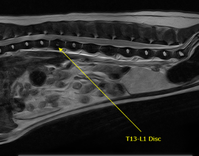
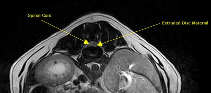
Nora is a five year old female spayed Bichon that presented for acute onset of rear limb weakness and back pain. A T3-L3 myelopathy (spinal cord problem) was suspected based on her neurologic exam and an MRI was performed. On the MRI you can see loss of the normally bright (hyperintense) inner disc (nucleus pulposus) at the T13-L1 disc space and the appearance of extradural (outside of the spinal cord) material above (dorsal) that disc space. On the axial image you can see extruded disc material on the left side of the spinal cord causing moderate extradural spinal cord compression. A left T13-L1 hemilaminectomy (decompressive spinal cord surgery) was performed immediately following the MRI. Nora did very well and went home with her family a couple days later!