Magnetic Resonance Imaging (MRI) is a powerful diagnostic tool that has revolutionized how we detect and treat neurologic disease in veterinary medicine. Prior to MRI, veterinarians were limited to radiographs, computed tomography (CT), myelograms, and other diagnostic tools. MRI is considered superior to these other modalities for evaluation of the brain and spinal cord due the ability of MRI to detect soft tissue pathology affecting the nervous tissue encased within the skull and vertebral canal.
MRI utilizes a strong magnet that causes protons within the patient to align with the magnet’s magnetic field. When a radiofrequency pulse is applied to the patient, those protons can be knocked out of alignment until the radiofrequency pulse is turned off. The amount of time it takes for realignment, and the energy released during realignment of those protons, can be detected by special coils/receivers. Based on the type of tissue (environment of those protons), and presence/absence of disease, the time of realignment and energy released can vary. Images can be created from this data, giving doctors a better idea of whether there is pathology affecting the CNS.
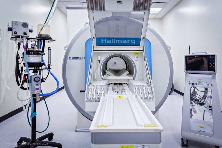
Wisconsin Veterinary Neurology and Surgical Center uses a 1.5 tesla (T) high field MRI just like is used on people. The biggest difference is that our patients need to be anesthetized to prevent motion during the scan. We utilize an MRI made by Hallmarq Advanced Veterinary Imaging, which is manufactured specifically for small animal patients. The use of a veterinary specific MRI has several advantages including small animal specific coils that achieve a much more accurate fit to veterinary anatomy, and a support team that understands the unique requirements of veterinary imaging.
WVNSC utilizes Hallmarq’s 3rd generation MRI. The generation is unique in that it uses zero helium.