We will be closed for emergencies & appts from 12/21-12/27 • We will be open for appts 12/28-12/29 • We will be closed for appts 1/1/25

Types of Intervertebral Disc Extrusions/Protrusion in the Cat and Dog

Adam Moeser, DVM, DACVIM (neurology) • November 4, 2024 Myelopathies caused by intervertebral disc disease are a very common reason that cat and dog owners seek veterinary care for their pets.  Intervertebral disc disease, however, is not a single disease process and there are various types of disc extrusions/protrusions.  The most well-known causes are Type I and Type II disc extrusions, but other diagnosis include a hydrated nucleus pulposus extrusion, an acute non-compressive nucleus pulposus extrusion, and it’s even possible to link a fibrocartilaginous embolism to intervertebral disc disease. At Wisconsin Veterinary Neurology, we are comfortable treating all kinds of disc disease.  If you’d like to refer a case or just consult on a case, please don’t hesitate to contact us.

Hansen Type I and II Intervertebral Disc Extrusion/Protrusion

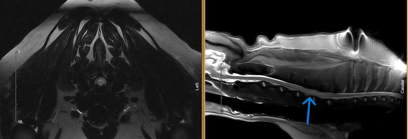
Figure 1: MRI images of a Hansen Type 1 Intervertebral Disc Extrusion.
*Figure 1: MRI images of a Hansen Type 1 Intervertebral Disc Extrusion. The T2 axial (left) and T2 sagittal (right) images show severe spinal cord compression due to a C2-C3 acute disc extrusion.

The most common type of disc problem in small animal veterinary medicine are Hansen Type I and II intervertebral disc extrusions/protrusions.  Type I disc extrusions involve extrusion of degenerative nucleus pulposus material into the spinal canal, which may cause compression of the spine and/or nerves.  This can result in signs ranging from pain to paralysis with loss of nociception.  This most commonly occurs in chondrodystrophic breeds (i.e. Dachshund, French Bulldog, Beagle, etc.) due to the premature chondroid degeneration of the nucleus pulposus.  This disc extrusion may occur anywhere along the spine, although usually not between T2 and T10 due to the intercapital ligament’s stabilizing properties in this region.  Cats usually suffer disc extrusions in the lumbar or lumbosacral region.  Treatment is via surgical decompression or conservative medical management.  When dogs are treated conservatively the prognosis ranges from 56-80% when sensation is intact, and poor (22%) when nociception is absent1.  Surgery is associated with an excellent prognosis (93-98%) when nociception is intact, and guarded (61%) when nociception is absent.

Type II Disc Disease involves degeneration and bulging of the annulus fibrosus, which can lead to compression of nervous tissues in the spinal canal.  This condition usually affects older non chondrodystrophic breeds such as German Shepherds and Labrador Retrievers.  While symptoms can vary just like with Type I disc extrusions, these patients are usually not very painful as this is more of a chronic disorder.  Treatment may be surgical or conservative, but the prognosis with surgery is more guarded compared to with Type I extrusions.

Hydrated Nucleus Pulposus Extrusion

A Hydrated Nucleus Pulposus Extrusion is a type of disc disease that usually affects the cervical spine in dogs, but can occur elsewhere.  I have not seen this diagnosis is cats, only dogs.  Some veterinarians know this problem as a “discal cyst”, but a more appropriate term is thought to be hydrated nucleus pulposus extrusion due to the lack of histologic evidence of a true cyst wall when samples have been submitted for pathologic review.  This problem tends to affect normal appearing discs (no evidence of chondroid degeneration), and on axial T2 MRI images the compression lies ventral to the spinal cord in a symmetrical distribution that some call the “seagull” sign.  When surgery is performed, a gelatinous fluid can be easily removed from the canal as soon as the dorsal longitudinal ligament is cut away.  Like all disc disease, the presence of nociception is a positive prognostic indicator.  The only study comparing surgery and conservative management showed a good/excellent prognosis with both2.

Acute Non-Compressive Nucleus Pulposus Extrusion (ANNPE)

An acute non-compressive nucleus pulposus extrusion has many other names, including: traumatic disc, missile lesion, and Type III disc herniation.  Regardless the name, this diagnosis involves the acute herniation of normal nucleus pulposus material into the canal at a high velocity.  This material strikes the spinal cord causing a concussive injury to the cord, but the amount of extradural compression is typically minimal.  Due to the minimal spinal cord compression, this is not a surgical diagnosis; conservative management using anti-inflammatory medications, pain management, and rest are all that is needed to treat this disease.  I usually recommend physiotherapy in addition to the medications and rest.  This diagnosis is made in both cats and dogs, and prognosis is associated with the degree of neurologic deficits at the time of diagnosis.  In dogs, one study found that when nociception is present at the time of diagnosis, 73% of patients will walk again3.  In that same study all dogs walked again when motor function was present at the time of diagnosis.  In cats, a 90% success rate has been reported4.  These patients often present identical to an FCE patient (non-painful, worse on one side), although they may display some signs of vertebral pain on palpation.
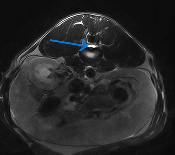
Figure 2: MRI image of a hydrated nucleus pulposus extrusion in a dog.
*Figure 2: MRI image of a hydrated nucleus pulposus extrusion in a dog. A T2 axial image showing a bilobed uniform brightness/hyperintensity underneath the spinal cord causing severe spinal cord compression.
*Figure 3. MRI images of an acute non-compressive nucleus pulposus extrusion in a dog.
*Figure 3. MRI images of an acute non-compressive nucleus pulposus extrusion in a dog. T2 sagittal (left) and axial (right) images showing loss of the normal volume of bright/hyperintense nucleus pulposus, and intramedullary hyperintensity of the cord above that same disc

Fibrocartilagenous Embolism

A fibrocartilaginous embolism (FCE) occurs when a piece of nucleus pulposus finds its way into the spinal cord arterial system causing an obstruction of blood flow to that area of the spine.  This disorder affects both dogs and cats.  Symptoms typically occur during high-impact athletic movements and are typically worse on one side.  These patients are typically non-painful, although they may appear very anxious/stressed at presentation.  The symptoms come on acutely, and usually after a couple hours these patients stabilize and don’t progress/worsen.  These patients are treated with good nursing care, rest, and physiotherapy. Anti-inflammatory medications have not been shown to help these patients and they are not painful.  I usually prescribe crate rest, trazodone, and recommend scheduling with a rehab certified veterinarian.  When nociception is intact the prognosis for recovering the ability to walk unassisted is good, with 85% of dogs having a good prognosis in one review of 393 cases5.  In that same study, only 10% of dogs had a good outcome if nociception was absent at the time of diagnosis.
*Figure 4: MRI images of a fibrocartilageous embolism in a dog.
*Figure 4: MRI images of a fibrocartilageous embolism in a dog. A T2 axial (left) and T2 STIR sagittal (right) show an intramedullary T2 hyperintensity within the spinal cord.

References

  1. Olby, N.J., da Costa, R.C., Levine, J.M., Stein, V. (2020), Prognosis Factors in Canine Acute Intervertebral Disc Disease. Front Vet Sci, 7.
  2. Nessler J,  Flieshardt C,  Tünsmeyer J,  Dening R,  Tipold A.  (2018), Comparison of surgical and conservative treatment of hydrated nucleus pulposus extrusion in dogs. J Vet Intern Med; 32: 1989–1995.
  3. De Risio, L., Adams, V., Dennis, R., and McConnell, F.J. (2009), Association of clinical and magnetic resonance imaging findings with outcome in dogs with presumptive acute noncompressive nucleus pulposus extrusion: 42 cases (2000-2007). J Am Vet Med Assoc, 234 (4): 495-504.
  4. Taylor-Brown FE, De Decker S. (2017), Presumptive acute non-compressive nucleus pulposus extrusion in 11 cats: clinical features, diagnostic imaging findings, treatment and outcome. Journal of Feline Medicine and Surgery,19(1):21-26.
  5. Bartholomew, K.A., Stover, K.E., Olby, N.J. and Moore, S.A. (2016), Clinical characteristics of canine fibrocartilaginous embolic myelopathy (FCE): a systematic review of 393 cases (1973–2013). Veterinary Record, 179: 650-650.