Hansen Type I and II Intervertebral Disc Extrusion/Protrusion

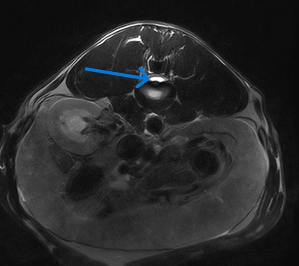
The most common type of disc problem in small animal veterinary medicine are Hansen Type I and II intervertebral disc extrusions/protrusions. Type I disc extrusions involve extrusion of degenerative nucleus pulposus material into the spinal canal, which may cause compression of the spine and/or nerves. This can result in signs ranging from pain to paralysis with loss of nociception. This most commonly occurs in chondrodystrophic breeds (i.e. Dachshund, French Bulldog, Beagle, etc.) due to the premature chondroid degeneration of the nucleus pulposus. This disc extrusion may occur anywhere along the spine, although usually not between T2 and T10 due to the intercapital ligament’s stabilizing properties in this region. Cats usually suffer disc extrusions in the lumbar or lumbosacral region. Treatment is via surgical decompression or conservative medical management. When dogs are treated conservatively the prognosis ranges from 56-80% when sensation is intact, and poor (22%) when nociception is absent1. Surgery is associated with an excellent prognosis (93-98%) when nociception is intact, and guarded (61%) when nociception is absent.
Type II Disc Disease involves degeneration and bulging of the annulus fibrosus, which can lead to compression of nervous tissues in the spinal canal. This condition usually affects older non chondrodystrophic breeds such as German Shepherds and Labrador Retrievers. While symptoms can vary just like with Type I disc extrusions, these patients are usually not very painful as this is more of a chronic disorder. Treatment may be surgical or conservative, but the prognosis with surgery is more guarded compared to with Type I extrusions.
Hydrated Nucleus Pulposus Extrusion
Acute Non-Compressive Nucleus Pulposus Extrusion (ANNPE)
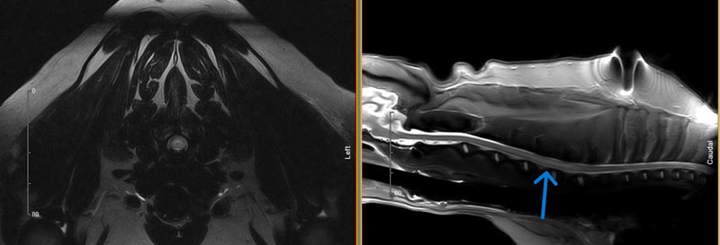
An acute non-compressive nucleus pulposus extrusion has many other names, including: traumatic disc, missile lesion, and Type III disc herniation. Regardless the name, this diagnosis involves the acute herniation of normal nucleus pulposus material into the canal at a high velocity. This material strikes the spinal cord causing a concussive injury to the cord, but the amount of extradural compression is typically minimal. Due to the minimal spinal cord compression, this is not a surgical diagnosis; conservative management using anti-inflammatory medications, pain management, and rest are all that is needed to treat this disease. I usually recommend physiotherapy in addition to the medications and rest. This diagnosis is made in both cats and dogs, and prognosis is associated with the degree of neurologic deficits at the time of diagnosis. In dogs, one study found that when nociception is present at the time of diagnosis, 73% of patients will walk again3. In that same study all dogs walked again when motor function was present at the time of diagnosis. In cats, a 90% success rate has been reported4. These patients often present identical to an FCE patient (non-painful, worse on one side), although they may display some signs of vertebral pain on palpation.


Fibrocartilagenous Embolism

References
- Olby, N.J., da Costa, R.C., Levine, J.M., Stein, V. (2020), Prognosis Factors in Canine Acute Intervertebral Disc Disease. Front Vet Sci, 7.
- Nessler J, Flieshardt C, Tünsmeyer J, Dening R, Tipold A. (2018), Comparison of surgical and conservative treatment of hydrated nucleus pulposus extrusion in dogs. J Vet Intern Med; 32: 1989–1995.
- De Risio, L., Adams, V., Dennis, R., and McConnell, F.J. (2009), Association of clinical and magnetic resonance imaging findings with outcome in dogs with presumptive acute noncompressive nucleus pulposus extrusion: 42 cases (2000-2007). J Am Vet Med Assoc, 234 (4): 495-504.
- Taylor-Brown FE, De Decker S. (2017), Presumptive acute non-compressive nucleus pulposus extrusion in 11 cats: clinical features, diagnostic imaging findings, treatment and outcome. Journal of Feline Medicine and Surgery,19(1):21-26.
- Bartholomew, K.A., Stover, K.E., Olby, N.J. and Moore, S.A. (2016), Clinical characteristics of canine fibrocartilaginous embolic myelopathy (FCE): a systematic review of 393 cases (1973–2013). Veterinary Record, 179: 650-650.
