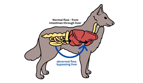
A not so uncommon cause of neurologic symptoms in young animals (puppies, kittens) is hepatic encephalopathy due to a portosystemic shunt. A portosystemic shunt is when the venous blood coming from the intestines bypasses the liver through an abnormal connection (shunt) between the portal vein and systemic circulation.
During fetal development the oxygenated placental blood bypasses the liver through connections between the portal vasculature and the fetal systemic vasculature. Shortly after birth (usually by 10 days), these connections close down and blood from the intestines first passes through the liver before reaching the systemic vasculature. In some young patients, one or multiple of these connections remain open, allowing blood to bypass the liver. Toxins are then allowed access to the Central Nervous System (CNS), which can cause a hepatic encephalopathy. Neurologic symptoms may include altered mentation (not as bright as usual), weakness, seizures, etc. The neurologic abnormalities are usually symmetrical, meaning that both sides of the body are affected equally. Affected dogs and cats may also be small in stature, and sometimes owners notice that neurologic symptoms worsen after eating.
Prior to performing an expensive diagnostic test like an MRI, we always recommend performing liver function testing on young animals (i.e. less than one year old) with neurologic abnormalities.
Liver function may be assesed via various laboratory diagnostics. If the total bilirubin is elevated, one potential cause is abnormal liver function. Total bilirubin is measured on most chemistry panels. Ammonia level and bile acids are other tests that are routinely performed in order to test liver function. Bile acids are the gold standard, and a pre/post prandial bile acids level should be selected to evaluate liver function whenever possible. Sometimes we don’t have time to wait for test results prior to performing an MRI, or the patient is too small to get two blood samples for pre and post prandial testing, and in these circumstances an ammonia level may be preferred.
If a liver shunt is suspected, the patient will be referred to an internal medicine specialist or radiologist for imaging of the liver and its associated vasculature. This may involve an abdominal ultrasound, but a CT portogram is the gold standard for diagnosing a portosystemic liver shunt.
Treatment of a liver shunt may involve medical treatments such as lactulose, antibiotics (amoxicillin, metronidazole), and a low protein diet. If surgery is possible (extrahepatic shunt), this is the treatment of choice as patients treated medically may still progress to liver failure.

