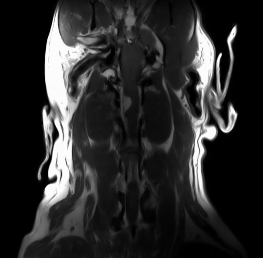
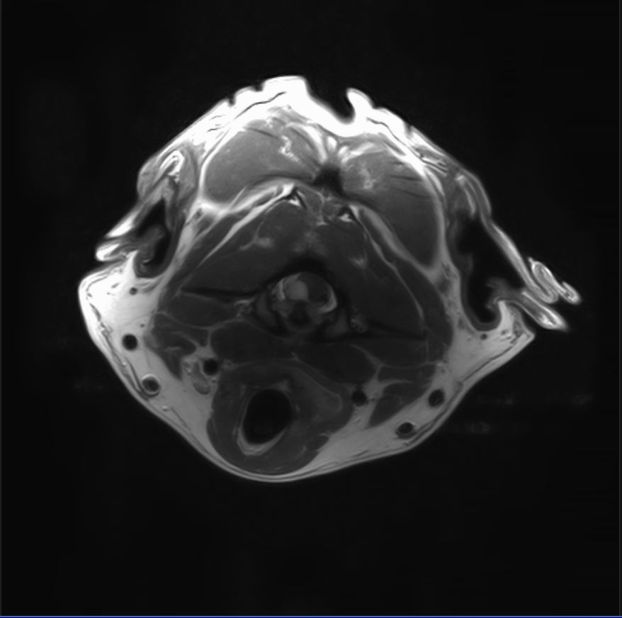
Hank is a nine-year-old male neutered Rhodesian Ridgeback who presented with signs of pain. Dr. Moeser performed an initial neurological exam and determined Hank’s pain to be emanating from his neck. An MRI identified a C1-C2 right-sided intradural extramedullary contrast enhancing mass. You can see it in the initial MRI images, below; there is a bright/white mass pushing on the right side of the spinal cord. Historically, dogs tend to get meningiomas in this region of the spine. In order to get a definitive diagnosis and make Hank feel better, Dr. Moeser surgically debulked the tumor. Post-surgical histopathology confirmed the mass was a meningioma.

This is a T1 post-contrast coronal MRI image of the cervical spine showing a contrast enhancing mass on the right side of the spine.

This is a T1 post-contrast axial MRI image of the cervical spine showing a contrast enhancing mass on the right side of the spine.

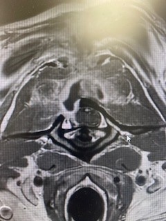
On the right, is a T1 post-contrast axial MRI image of the cervical spine showing the previous surgical site where the bone has been removed. There is still contrast enhancing tissue, but the majority of the tumor has been removed.
Post surgery, Hank received 4 weeks/20 sessions of low-dose radiation treatment at the University of Wisconsin-Madison Veterinary Oncology Department. While the meningioma will regrow, it will likely regrow very slowly. Hank is living his best life and it is possible he will never experience symptoms from the mass again.
Hank’s journey is a testament to the power of expert care and unwavering determination. If your pet is facing neurological challenges, you’re not alone. We’re here to help.
We specialize in diagnosing and treating neurological conditions in pets like Hank, helping them regain mobility and quality of life. Contact us to schedule a consultation and see how we can support your pet’s journey to recovery.

